We makethe bodymeasurable.
A San Diego laboratory where cardiovascular and metabolic devices are invented, proven in large animals, and built into companies. Eleven engineers, clinicians and technicians. One building. Bench to first-in-human without a vendor contract.
San Diego, California
Est. 2007
200+
Issued and pending patents
Conductance, retroperfusion, pericardial access, magnetic occlusion
5
Portfolio companies
Each built on a platform proven in the laboratory downstairs
300+
Full-length publications
From the founder's laboratory, 373 of them indexed in PubMed
2025
First FDA clearance
Fecobionics Anorectal System, K242666, cleared February 2025
Everything here descends
from one measurement.
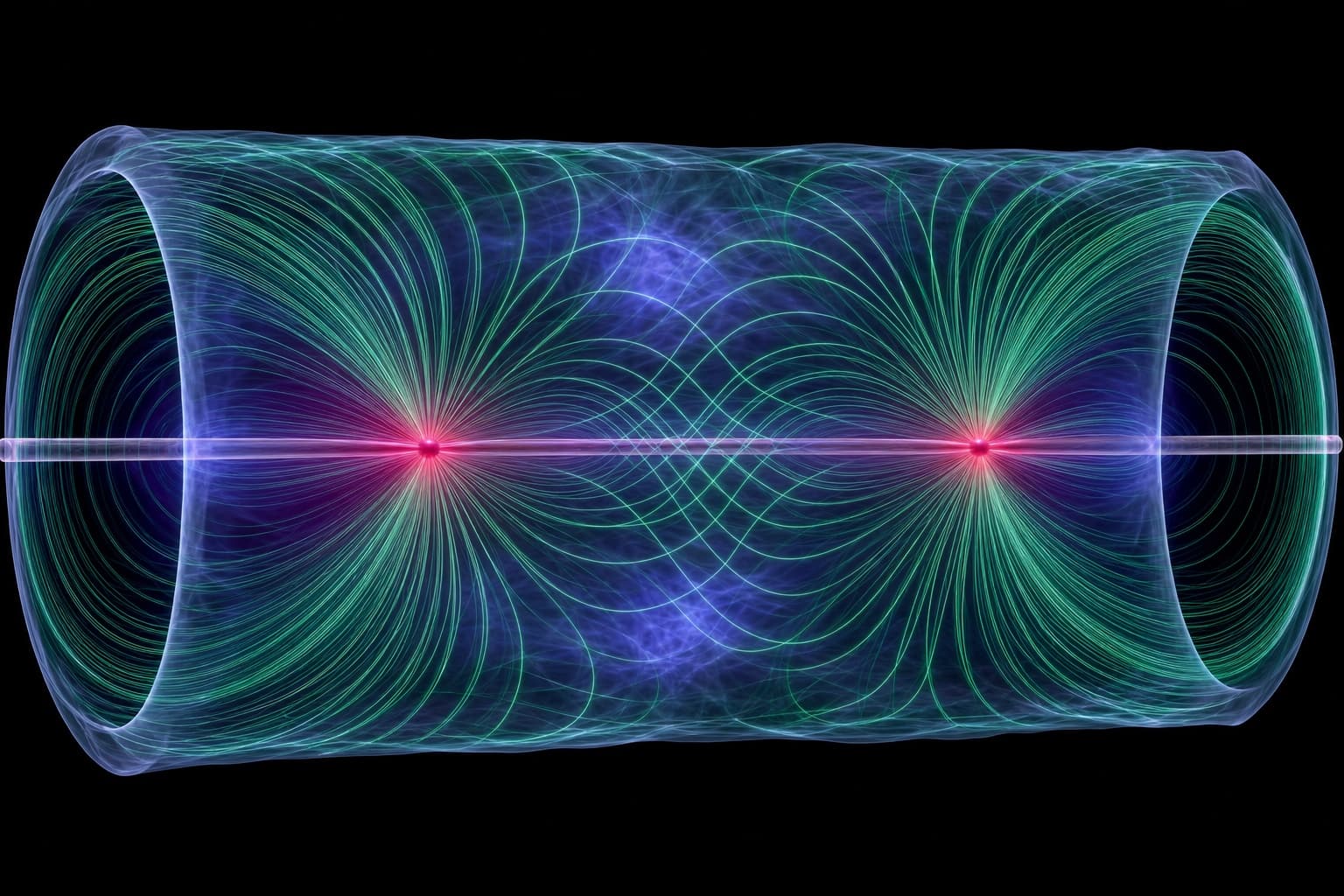
Nearly everything here descends from one measurement. Pass a small current between two electrodes inside a vessel and the tissue answers. The answer is geometry: the exact cross-section of the lumen you are standing in, live, without contrast and without guesswork.
Drag along the vessel to move the catheter. Arrow keys work too.
Lumen area
7.65mm²
Diameter
3.12mm
Conductance
5.35mS
Area stenosis
0%
Inject
Two excitation electrodes on the catheter shaft drive a low-amplitude alternating current, tens of microamps at tens of kilohertz, through blood and vessel wall.
Sense
Two detection electrodes sit a millimeter apart between them and read the voltage that survives. Current divided by voltage is conductance, and conductance scales with the area the current had to cross.
Separate
Some of that current leaks into the wall and the tissue beyond it. Two injections at different saline concentrations cancel the leak and leave the lumen alone. It is a novel approach to a correction the field has used since the 1980s, and it is what makes the number trustworthy.
Act
The interventionalist now sizes a stent, a valve annulus or a balloon against a measured area rather than an estimate read off a shadow.
Four problems, each
measured before it is treated.
Every program starts with a quantity a clinician cannot currently see, and ends with a device that reports it during the procedure rather than after it.
5 companies, each
proven in this building.
An incubator that owns an operating room does not have to take a founder's word for whether the device works. Nothing is spun out of 3DT until it has survived a large-animal study downstairs.
- 01
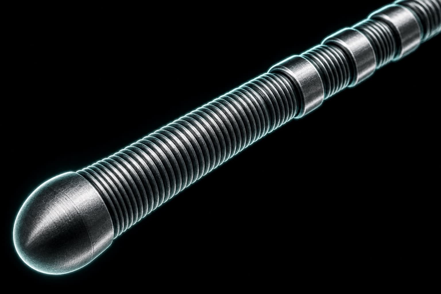
Intelligent Delivery Systems
Guidewires that measure.
Coronary and peripheral intervention
- 02
GRest
Bariatric outcomes without the amputation.
Metabolic and bariatric
- 03
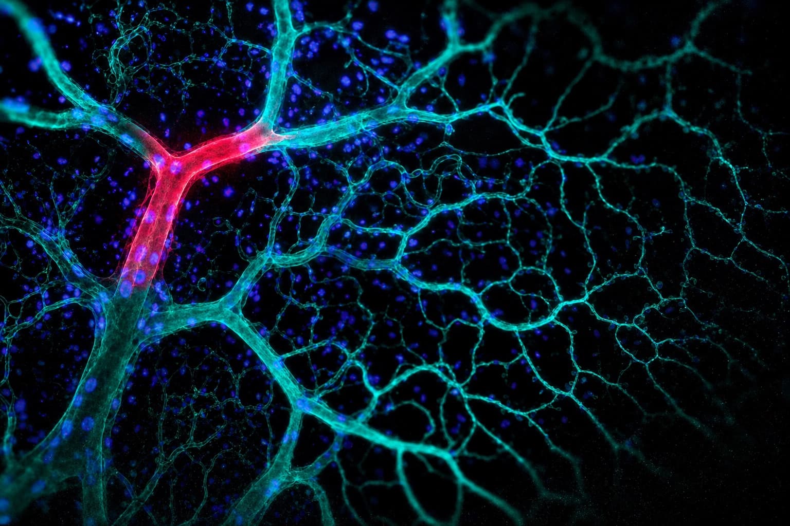
Retroperfusion
Flow, running backward.
Circulatory support
- 04
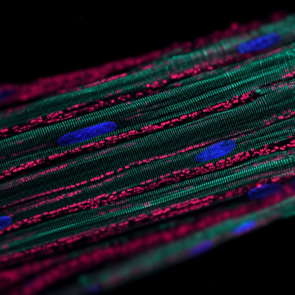
Pericardial Access
A door in the heart wall.
Cardiac delivery
- 05
GI Bionics
Fecobionics.
Gastroenterology
Bench to first-in-human,
without leaving the building.
Most device companies rent their evidence. They send a prototype to one vendor for bench testing, another for the animal study, a third for histology, and wait weeks between answers. Here all of it sits on one corridor, which is why an iteration takes days.
Surgical
- Two sterile surgical suites with wide-screen viewing displays
- Post-surgical recovery rooms
- Anesthesia monitoring and extended physiological monitoring
Imaging
- Two flat-panel C-arms in a poly-diagnostic configuration
- Multi-angle acquisition with DICOM archiving
- Intravascular ultrasound (IVUS)
- Trans-thoracic and trans-esophageal echocardiography
- Peripheral vascular Doppler ultrasound
Pathology
- Experimental Pathology and Immunopathology Laboratory (EPIL)
- Immunofluorescence and immunoperoxidase
- Single, double and triple-antibody methods
- Led by a board-certified pathologist
Computational
- Computational fluid dynamics and finite element analysis
- Fluid-structure interaction
- Mass and heat transport, electromagnetic simulation
- High-performance computing through university partners
All three are held by the California Medical Innovations Institute, the non-profit research institute in the same building with which 3DT shares a founder. Animal work runs in its facility and under its IACUC.